Lab News
 May, 2016:
Edmund Hollis 2nd, Nao Ishiko, Ting Yu , Chin-Chun Lu, Ariela Haimovich, Kristine Tolentino, Alisha Richman, Anna Tury, Shih-Hsiu Wang, Maysam Pessian, Euna Jo, Alex Kolodkin and Yimin Zou. Ryk controls remapping of motor cortex during functional recovery after spinal cord injury. Nature Neurosci. 2016 May;19(5):697-705
Please click here to read the news.
 Apr 11, 2016:
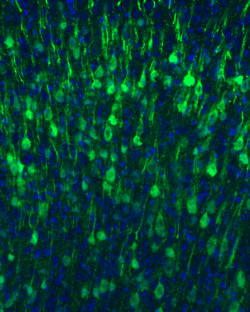
Hollis et al showed that conditionally knocking out Ryk led to increased corticospinal axon plasticity and functional recovery. Motor cortex reorganized such that the hindlimb cortex controls the forelimb with continued forelimb reaching task training:
Please click here to read the news.
Jan 19, 2015:
Hollis et al showed that reorganization of synaptic contacts can strengthen the spared circuits and lead to functional recovery after partial spinal cord injury. And blocking repulsive Wnt signalling increases axon plasticity and synaptic connections that drive greater functional recovery.Please click here to download the PDF file.
Dec 23, 2014:
Hollis et al reported a more robust conditioning lesion model. Injection of a chemical demyelinating agent, ethidium bromide, into the sciatic nerve induces a similar set of regeneration-associated genes and promotes a 2.7-fold greater extent of sensory axon regeneration in the spinal cord than sciatic nerve crush. This study provides a new method for investigating the underlying mechanisms of the conditioning response and suggests that loss of the peripheral myelin maybe a major signal to change the intrinsic growth state of adult sensory neurons and promote regeneration.Please click here to download the PDF file.
Dec 4, 2013:
Onishi et al published new findings on signaling mechanisms that mediates growth cone turning. Commissural axons engage planar cell polarity (PCP) signaling components to turn in a Wnt gradient. Frizzled3, a Wnt receptor, undergoes endocytosis via filopodia tips. Wnt5a increases Frizzled3 endocytosis, which correlates with filopodia elongation. He discovered an unexpected antagonism between Dishevelleds, which may function as a signal amplification mechanism in filopodia.Please click here to download the PDF file.
Oct 7, 2013:
Anna Tury et al published their studies on changes of Wnt signaling components in an ALS mouse model. Amyotrophic lateral sclerosis (ALS) is a fatal neurodegenerative disease characterized by progressive paralysis due to the selective death of motor neurons of unknown causes. She focused on two non-canonical Wnt signaling components, atypical PKC (aPKC) and a Wnt receptor, Ryk. aPKC expression was increased in motor neurons of the lumbar spinal cord in SOD1 (G93A) mice at different stages and may be sequestered in SOD1 aggregates. Ryk expression was also increased in the motor neurons and the white matter in the ventral lumbar spinal cord of mutant SOD1 mice with a peak at early stage. These observations indicate that Wnt/aPKC and Wnt/Ryk signaling are altered in SOD1 (G93A) mice, suggesting that changed Wnt signaling may contribute to neurodegeneration in ALS.Please click here to download the PDF file.
August 17, 2012:
Work done by Edmund Hollis II on how Wnt-Ryk signaling limits sensory axon regeneration following spinal cord injury was published in PNAS.
Conditioning lesion of the peripheral branch of dorsal column axons is a well-known paradigm enabling the central branch to regenerate after injury to the spinal cord. However, only a small number of regenerating axons enter grafted substrates, and they do not grow beyond the lesion. We found that conditioning lesion induces, in addition to growth-stimulating genes, related to receptor tyrosine kinase (Ryk), a potent repulsive receptor for Wnts. Wnts are expressed around the site of spinal cord injury, and we found that grafted bone marrow stromal cells secreting the Wnt inhibitors secreted frizzled-related protein 2 or Wnt inhibitory factor 1 enhanced regeneration of the central branch after peripheral conditioning lesion. Furthermore, we found that Wnt4-expressing grafts caused dramatic long-range retraction of the injured central branch of conditioned dorsal root ganglion neurons. Macrophages accumulate along the path of receding axons but not around Wnt4-expressing cells, suggesting that the retraction of dorsal column axons is not a secondary effect of increased macrophages attracted by Wnt4. Therefore, Wnt-Ryk signaling is an inhibitory force co-induced with growth-stimulating factors after conditioning lesion. Overcoming Wnt inhibition may further enhance therapies being designed on the basis of the conditioning-lesion paradigm.
Please click here to download the PDF file.
March 1, 2011:
Sonal Thakar joined the lab to study the mechanisms underlying the establishment of neural circuits in the visual system. Sonal obtained Ph.D. from UT Southwestern and worked with Mark Henkemeyer on the roles of ephinB/EphB signaling system in retiontopic mapping.
February 15, 2011:
Work by Beth Shafer, Keisuke Onishi, Charles Lo and Gulsen Colakoglu on how planar cell polarity signaling components mediate Wnt responses in commissural axon growth cones was published in Developmental Cell.
Summary:
Although a growing body of evidence supports that Wnt-Frizzled signaling controls axon guidance from vertebrates to worms, whether and how this is mediated by planar cell polarity (PCP) signaling remain elusive. We show here that the core PCP components are required for Wnt5a-stimulated outgrowth and anterior-posterior guidance of commissural axons. Dishevelled1 can inhibit PCP signaling by increasing hyperphosphorylation of Frizzled3 and preventing its internalization. Vangl2 antagonizes that by reducing Frizzled3 phosphorylation and promotes
its internalization. In commissural axon growth cones, Vangl2 is predominantly localized on the plasma membrane and is highly enriched on the tips of the filopodia as well as in patches of membrane where new filopodia emerge. Taken together, we propose that the antagonistic functions of Vangl2 and Dvl1 (over Frizzled3 hyperphosphorylation and endocytosis) allow sharpening of PCP signaling locally on the tips of the filopodia to sense directional
cues, Wnts, eventually causing turning of growthcones.
Please click here to download the PDF and here to see a news article related to this story.
December 15, 2010:
Dr. Liqing Wang, who worked on spinal cord commissural axon guidance with Dr. Sun On Chan at the Chinese University of Hong Kong, joined the lab. Liqing will study how molecular genetic programs specify neuronal wiring diagrams.
November 24, 2010:
Work by a former graduate student, Ali Fenstermaker, in collaboration with Jeroen Pasterkamp's lab, was published in Journal of Neuroscience: "Wnt/Planar Cell Polarity Signaling Controls the Anterior-Posterior Organization of Monoaminergic Axons in the Brainstem". Click here to download the PDF of this paper.
Abstract:
Monoaminergic neurons [serotonergic (5-HT) and dopaminergic (mdDA)] in the brainstem project axons along the anterior-posterior axis. Despite their important physiological functions and implication in disease, the molecular mechanisms that dictate the formation of these projections along the anterior-posterior axis remain unknown. Here we reveal a novel requirement for Wnt/planar cell polarity signaling in the anterior-posterior organization of the monoaminergic system. We find that 5-HT and mdDA axons express the core planar cell polarity components Frizzled3, Celsr3, and Vangl2. In addition, monoaminergic projections show anterior-posterior guidance defects in Frizzled3, Celsr3, and Vangl2 mutant mice. The only known ligands for planar cell polarity signaling are Wnt proteins. In culture, Wnt5a attracts 5-HT but repels mdDA axons, and Wnt7b attracts mdDA axons. However, mdDA axons from Frizzled3 mutant mice are unresponsive to Wnt5a and Wnt7b. Both Wnts are expressed in gradients along the anterior-posterior axis, consistent with their role as directional cues. Finally, Wnt5a mutants show transient anterior-posterior guidance defects in mdDA projections. Furthermore, we observe during development that the cell bodies of migrating descending 5-HT neurons eventually reorient along the direction of their axons. In Frizzled3 mutants, many 5-HT andmdDAneuron cell bodies are oriented abnormally along the direction of their aberrant axon projections. Overall, our data suggest that Wnt/planar cell polarity signaling may be a global anterior-posterior guidance mechanism that controls axonal and cellular organization beyond the spinal cord.

November 18, 2010:
Dr. Youjun Chen from Bill Snider's lab at University of North Carolina, Chapel Hill, paid a visit to the lab after the Society for Neuroscience Meeting and shared her elegant work on the role of APC in regulating neuronal morphology.
November 1, 2010:
Ali Fenstermaker, who just graduated in September, will join Joseph Gleeson's lab as a postdoctoral fellow starting in early 2011. We will miss you, Ali!
September 30, 2010:
Ali Fenstermaker successfully defended her thesis, Mechanisms of Axon Pathfinding and Survival. Congratulations to Ali!
February 26, 2010:
Beth Shafer successfully defended her doctorate thesis today. Her work on how Wnt signaling mechanisms in controlling growth cone navigation will help us understand how neural circuits are established. Congratulations, Beth!
December 24, 2009:
Liseth Parra's paper on Sonic Hedgehog inducing Semaphorin chemorepulsion was highlighted in a News and Views at Nature Neuroscience. Click here to read the article.
December 1, 2009:
Liseth Parra's paper was selected and evaluated by Faculty of 1000 Biology. Click here to view the evaluation.
November 29, 2009:
Work from a former graduate student, Liseth Parra, is now published online at Nature Neuroscience-- "Sonic hedgehog induces response of commissural axons to Semaphorin repulsion during midline crossing". Click here to download a PDF of this paper.
Abstract
Pathfinding axons change responses to guidance cues at intermediate targets. During midline crossing, spinal cord commissural axons acquire responsiveness to class 3 Semaphorins and Slits, which regulate their floor plate exit and restrict their post-crossing trajectory into a longitudinal pathway. We found that Sonic Hedgehog (Shh) could activate the repulsive response of pre-crossing axons to Semaphorins. Blocking Shh function with a monoclonal antibody to Shh, 5E1, in 'open-book' explants or by expressing a dominant-negative form of Patched-1, Ptch1Deltaloop2, or a Smoothened (Smo) shRNA construct in commissural neurons resulted in severe guidance defects, including stalling and knotting inside the floor plate, recrossing, randomized anterior-posterior projection and overshooting after crossing, reminiscent of Neuropilin-2 mutant embryos. Enhancing protein kinase A activity in pre-crossing axons diminished Shh-induced Semaphorin repulsion and caused profound midline stalling and overshooting/wandering of post-crossing axons. Therefore, a morphogen, Shh, can act as a switch of axon guidance responses.
November 10, 2009:
Elena Rubio de la Torre, PhD. is joining the lab as a postdoctoral fellow from Institude of Parasitology and Biomedicine "Lopez-Neyra", CSIC, Granada (Spain). Elena studied molecular pathogenesis of Parkinson's disease and will set up models for neurodegenerative disorders.
November 2, 2009:
Keisuke Onishi, who received his Ph.D. from Institute of Molecular and Cellular Biosciences at the University of Tokyo, is joining the lab at the beginning of 2010 as a postdoctoral fellow. He brings in expertise in cell polarity signaling in microtubule organization and dynamics.
August 28, 2009:
Congratulations to Beth Shafer on winning the Society for Neuroscience's Travel Award! Beth and Alisha will be attending the Society for Neuroscience conference in Chicago, IL.
June 25, 2009:
Liseth Parra successfully defends her thesis, Molecular Mechanisms of Axon Guidance in the Developing Spinal Cord, and receives her PhD! She is joining Dr. Filippo Rijili's lab as a postdoctoral fellow at the Friedrich Miescher Institute in Basel, Switzerland in fall 2009.

