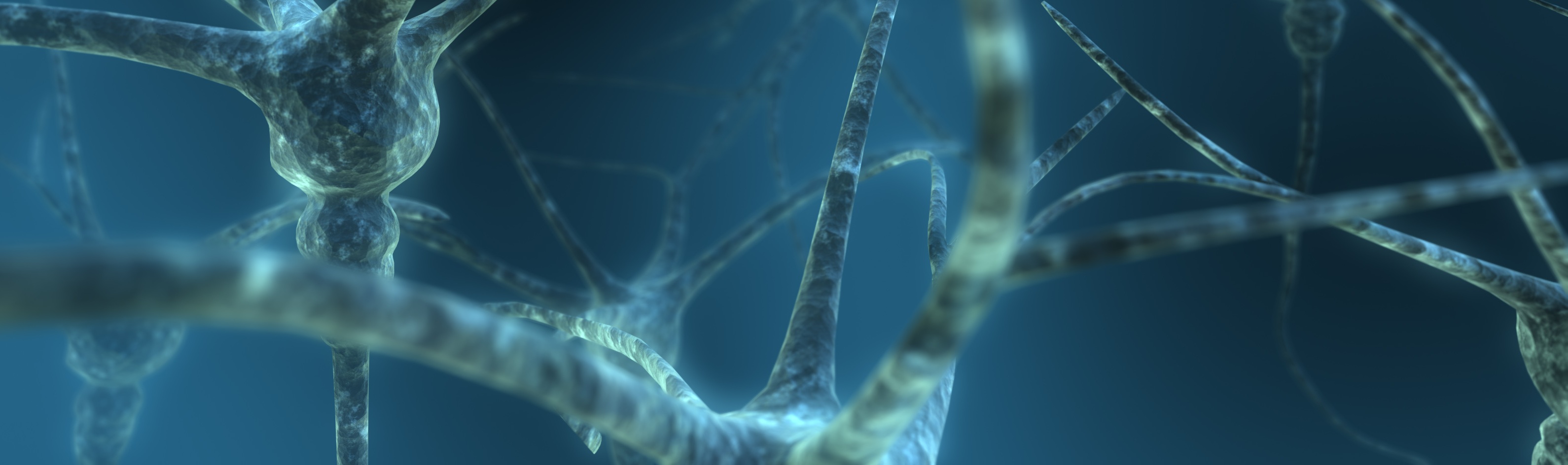
Normal behavioral functions rely on precise and complex neural circuits linking large ensembles of neurons. In development, axons and dendrites extend in a highly directed manner and elaborate intricate branches and arbors to establish organized patterns of synaptic connections. A genetic program plays a profound role in the formation of these networks by specifying neuronal cell types, positioning neurons, guiding axons and dendrites and forming appropriate functional synapses. Neural activity shapes circuit development and coordinate with the genetic program.
After the nervous system matures, certain areas of nervous system remain highly plastic in adulthood, whereas others are much less malleable. The regenerative ability of the adult central nervous system is generally highly limited. Abnormal circuit development and degenerative disorders lead to behavioral deficits.
We study development, degeneration and repair of neural circuits.
1. Molecular and Cellular Mechanisms of Axon Guidance

Guidance cues specifying axonal organization
Axonal connections are highly organized and precisely patterned. Much of the organization is achieved by the action of a large number of axon guidance cues, which control the direction of axon pathfinding and target selection in development. We are studying essential molecular determinants, particularly extracellular guidance cues, which control the directed pathfinding of axons and specify patterns of synaptic connections.
Using a number of axon model systems, spinal cord commissural neurons, dorsal root ganglion neurons, retinal ganglion cells, corticospinal tracts, and serotonergic and dopaminergic neurons in the brainstem, we are identifying guidance molecules and characterizing their roles in controlling the direction of growth in vivo. We study the function of several families of axon guidance molecules, including the Wnt family proteins, which are conserved directional cues.  Lyuksyutova et al. Liu et al. Fenstermaker et al Salinas and Zou Zou and Lyuksyutova Gradients of molecular guidance cues, such as the Wnt gradients, control the direction of axon navigation in vivo. Many gradients appear to be at least in part created by their graded gene expression across embryonic structures. We are interested in the mechanisms establishing and maintaining these gradients.
Signaling pathways that guide axons
The growth cone is a specialized structure localized at the tip of a growing axon responsible for sensing and responding to the guidance cues present in its micro-enviroment. Guidance molecules are detected by receptors on the surface of growth cones. Once the cues bind to their receptors, signals are then transduced across the plasma membrane and interpreted in the growth cones. We found that signaling pathways that specify the apical-basal and planar cell polarity of epithelial cells mediate Wnt signaling in axon guidance
Wolf et al.
Fenstermaker et al.
Shafer et al.
We are currently studying how these two cell polarity signaling pathways interact with each other in growth cone guidance.
Axons often face multiple guidance molecules and need to integrate signaling activities to make correct guidance decisions. For axons that travel a long distance, they often have complex trajectories that are made of segments punctuated by intermediate targets before they reach their final target area. Growth cones often stay at the intermediate targets for certain period of time, change their responsiveness and become sensitive to new guidance cues when they leave the intermediate targets.
We are studying how growth cones integrate signaling pathways activated by different guidance cues and how their sensitivity to guidance cues change while growing across intermediate targets. We found a classic morphogen, Sonic Hedgehog can switch on responsiveness of commissural axons to Semaphorin at the midline Parra and Zou
Cell biology of growth cone turning
For growth cones to turn in response to guidance molecules, certain directed or asymmetrical processes within the growth cone need to be engaged. It is well known that the cytoskeleton and the growth cone membrane undergo significant reorganization, presumably regulated by signals activated by guidance molecules. But little is known about how these changes cause growth cone turning. We are using the Wnt family guidance molecules as a model system to study the fundamental cellular machinery responsible for growth cone turning and to understand how this machinery responds to guidance cues. One approach we are taking is to identify the key downstream signaling components that regulate cytoskeleton and membrane trafficking, determine their localization and activation in the growth cone and study how these signal activations correlate with changes in cytoskeletal and membrane dynamics. For this, we are using live imaging techniques combined with biochemistry and molecular biology approaches. We found that planar cell polarity signaling component, Vangl2, is enriched in tips of growing filopodia but not in tips of shrinking filopodia, suggesting that the growth cone filopodia tips are the most sensitive part of the growth cone
Shafer et al.
2. Wiring for Function
The connections among many areas of the nervous system are highly specific and organized. Part of the organization is set up during pathfinding where axon-axon interactions self sort each other and directional cues lead them to proper target area. Once axons arrive at the target area, they need to establish various patterns of synaptic connections by seeking out correct post-synaptic partners.
One type of synaptic connection pattern is topographic connections, which convey smooth and continuous positional information between two connected areas. Our studies suggest that diffusible guidance cues, such as Wnts, also play a role in specifying topographic position by controlling axon target selection. We found that Wnt3 and EphrinB1 are two counterbalancing mapping forces in retinotectal projections along the dorsal-ventral (medial-lateral) axis.
Schmitt et al.
We are currently testing whether this "two-molecular-gradient model" is a general mechanism for topographic mapping and how two opposing mapping activities lay out topographic connections. Another common wiring strategy is laminar-specific targeting. For example, different retinal ganglion cell axons find their synaptic targets in different recipient layers in the optic tectum (in chick) and superior colliculus (in mammals). Layer-specific targeting contributes significantly to the specific patterns of synaptic connections and creates cellular and subcellular precisions, which are essential for encoding behavior. We are investigating the mechanisms of this level of brain wiring to understand how functional neural circuits emerge from the molecular cues and their potential interactions with neural activity in this process.
3. Injury and Regeneration of the Mammalian Central Nervous System

In the adult mammalian central nervous system axons generally do not naturally regenerate. We are interested in understanding the mechanisms regulating axon regrowth and regeneration and how these mechanisms could be used to repair the injured central nervous system.
Traumatic injury in the central nervous system leads to changes of gene expression in the injured areas as well as in neurons whose connections are altered. Some of these induced genes are important regulators of developmental processes, such as axon guidance. However, the roles of these reinduced developmental genes in injury response are unclear. We are studying the role of reinduced Wnt signaling system in injury response, including regulating axon regeneration.
Liu et al. The reinduec Wnt inhibitory system also limits sensory axon regeneration even after conditioning lesion, suggesting that Wnt-Ryk signaling is a general barrier for axon regeneration in the central nervous system.
Hollis et al.
Our final goal is to develop therapeutic approaches to promote regeneration and functional recovery after spinal cord injury.
4. Mechanisms of Neurodegenerative Disorders

Axon loss in degenerative disorders or traumatic injury leads to permanent changes/loss of circuits and function, making it challenging to improve function in affected patients. Because in the mammalian central nervous system axons generally do not naturally regenerate, another approach to try to prevent, or treat neurodegenerative disorders would be by protecting the existing neurons and axons, and impeding their degeneration. In this respect, we are studying the mechanisms that control axon stability and protection, as well as the ones that lead to axon degeneration in several neurodegenerative disease models, such as amyotrophic lateral sclerosis (ALS) and traumatic injury. Our aim is to understand the neurobiological mechanisms that regulate the stability of axons with the aim to develop new therapies for axon protection.